Radiation damage and helium buildup in structural nuclear materials
Generation IV nuclear power plants will expose the structural materials used in the reactor core to higher temperatures and faster neutron spectra than in previous reactor designs. This will give rise to complex effects in the damage microstructure of the structural materials. Furthermore, the ITER and DEMOnstration fusion reactors aim to demonstrate the commercial viability of fusion power and will expose the materials used in the core to even harsher thermal and radiation environments. Due to the varying neutron energy spectra across fission and fusion reactors, the amount of damage (measured in “displacements per atom” or “DPA”) the material will experience will vary. In addition to this, the rate of helium introduction within the materials (measured in “helium atomic parts per million” or “He-appm”) will also vary depending on the neutron spectra leading to a range of He-appm/DPA ratios. Helium has a low solubility in materials and generally agglomerates in areas of low electron density (e.g. vacancies and grain boundaries) and this has important consequences on the types of defects which are formed and how the materials properties degrade under these conditions.
Using the Microscopes and Ion Accelerators for Materials Investigations (MIAMI) facility, we examine the effects of varying the He-appm/DPA ratio and temperature on the damage microstructure of model structural nuclear materials such as iron-chrome and silicon carbide. This involves using helium ion irradiation at different energies between room temperature and 1300°C. The overall aim of this work is to build up a detailed dataset of defect microstructure of these materials as a function of He-appm/DPA ratio, temperature and total dose.
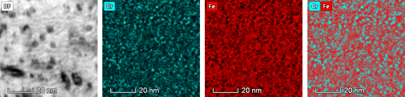 Scanning transmission electron micrograph and chemical maps of Cr separation in ferritic steel after irradiation with helium ions (Credit, A. Carruthers, University of Manchester/ RW Harrison, University of Huddersfield)
Recent Publications
Damage microstructure evolution of helium ion irradiated SiC under fusion relevant temperatures
Harrison, R. W., Ebert, S., Hinks, J. A. & Donnelly, S. E. 30 Apr 2018 In : Journal of the European Ceramic Society.
Influence of pre-implanted helium on dislocation loop type in tungsten under self-ion irradiation
Harrison, R., Hinks, J. & Donnelly, S. 20 Mar 2018 In : Scripta Materialia. 150, C, p. 61-65 5 p.
A study of the effect of helium concentration and displacement damage on the microstructure of helium ion irradiated tungsten
Harrison, R. W., Greaves, G., Hinks, J. A. & Donnelly, S. E. Nov 2017 In : Journal of Nuclear Materials. 495, p. 492-503 12 p.