MEIS Facility
The UK Medium Energy Ion Scattering facility, (MEIS) uses an ion beam probe to enable the analysis of the structure and properties of the top atomic layers of crystalline materials as well as the high-resolution depth profiling of non-crystalline layers of nano-meter thickness. The ion beam consists typically of hydrogen or helium ions which are accelerated to an energy between 100 keV and 200 keV and then scatter off the sample surface under investigation. After colliding with the surface atoms, the backscattered probe ions carry information about the surface layer in the energy and angular distributions of the backscattered ions which are analysed using an electrostatic energy analyser equipped with a 2D detector. Computer simulations and/or calculations are used for the extraction of the surface information. MEIS is a comparatively unique facility as there are fewer than ten MEIS machines world-wide.
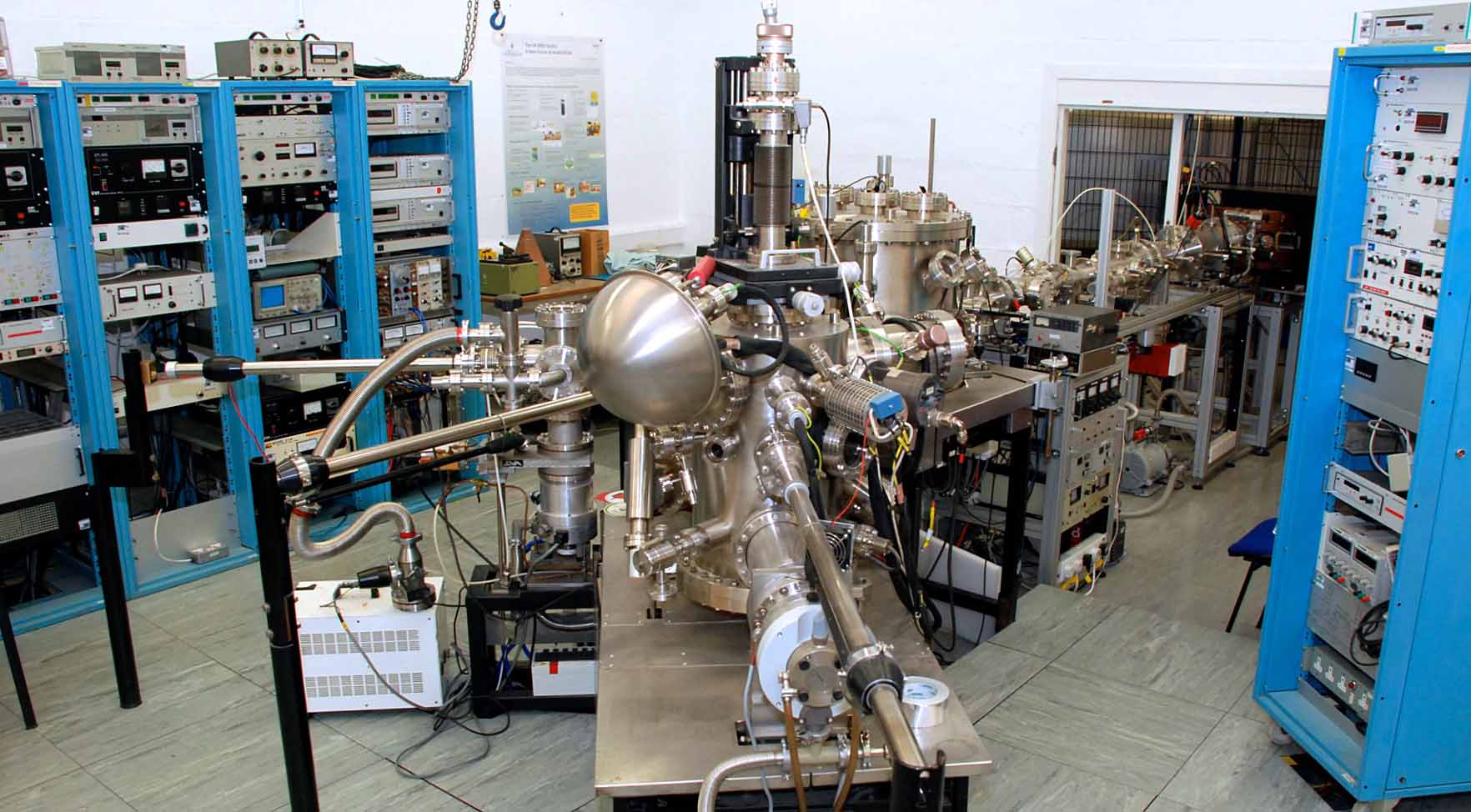
The MEIS experimental station comprises four interconnected UHV systems between which samples can be transferred under UHV conditions:
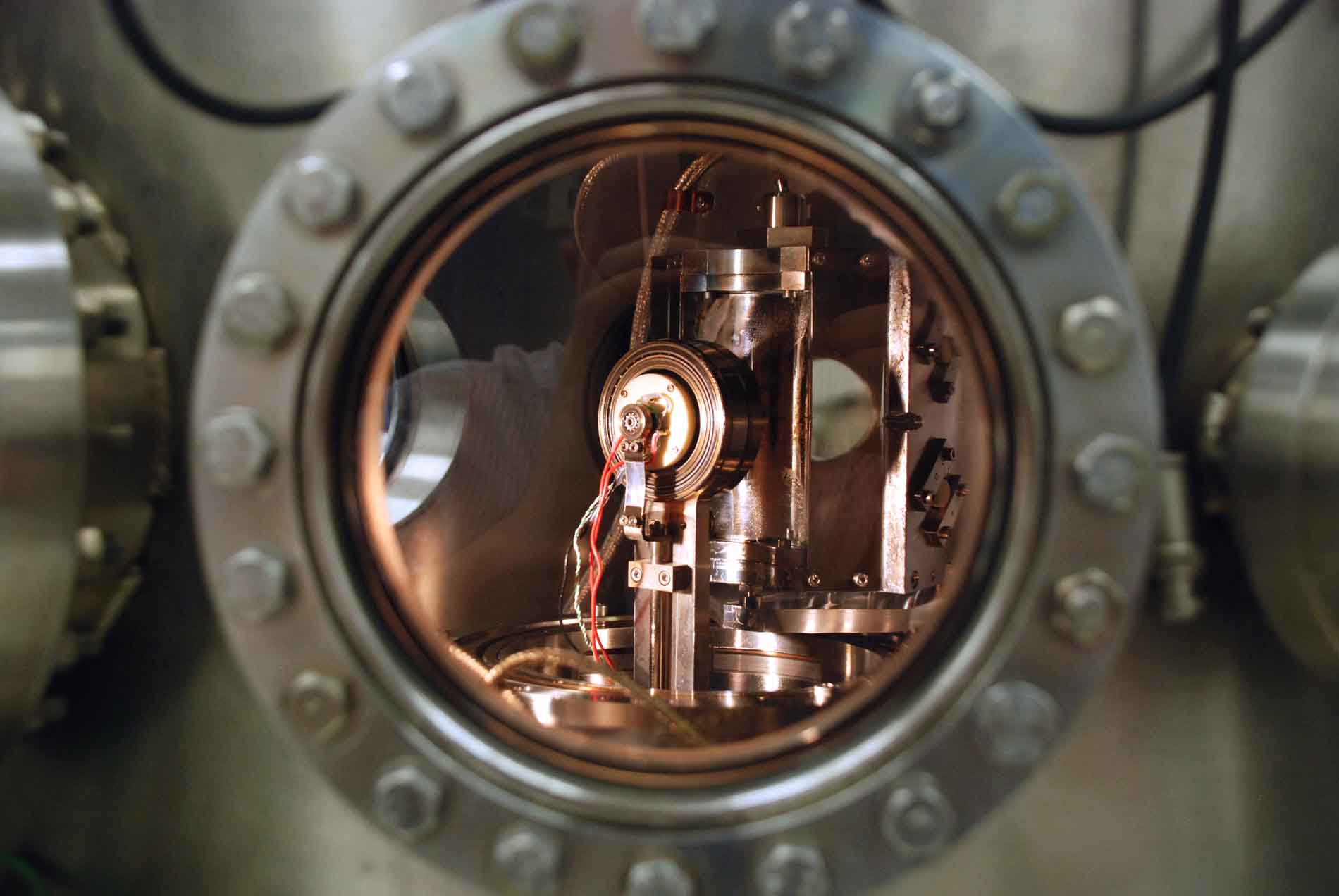
- The scattering chamber, in which the MEIS experiments are performed, houses the 3-axis sample goniometer, the electrostatic ion energy analyser and 2-dimensional (energy and angle) position sensitive detector.
- The preparation chamber, the facilities of which include LEED, Auger, sputter cleaning, evaporation sources and gas dosing is used for sample preparation and characterisation prior to ion scattering experiments.
- The storage chamber, which forms the junction of the sample transfer system, in which six samples can be stored.
- The loadlock which is a fast pump down chamber used for introducing samples into the vacuum. All 3 chambers are fitted with sample heating facilities.
We hope that the MEIS system will support a thriving user community. Potential users, for research and commercial applications are invited to contact either Professor Jaap Van Den Berg (J.VanDenBerg@hud.ac.uk) or Dr. Andrew Rossall (A.Rossall@hud.ac.uk) to discuss possibilities.
MEIS Facility
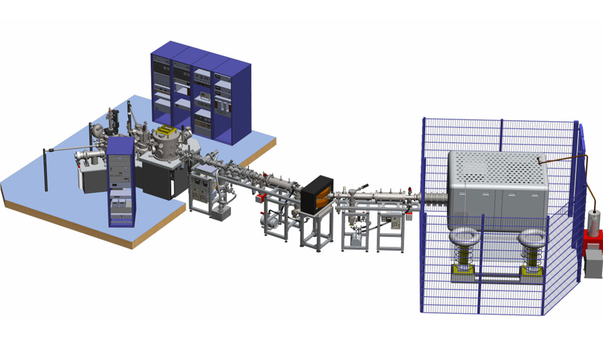
MEIS Facility plan
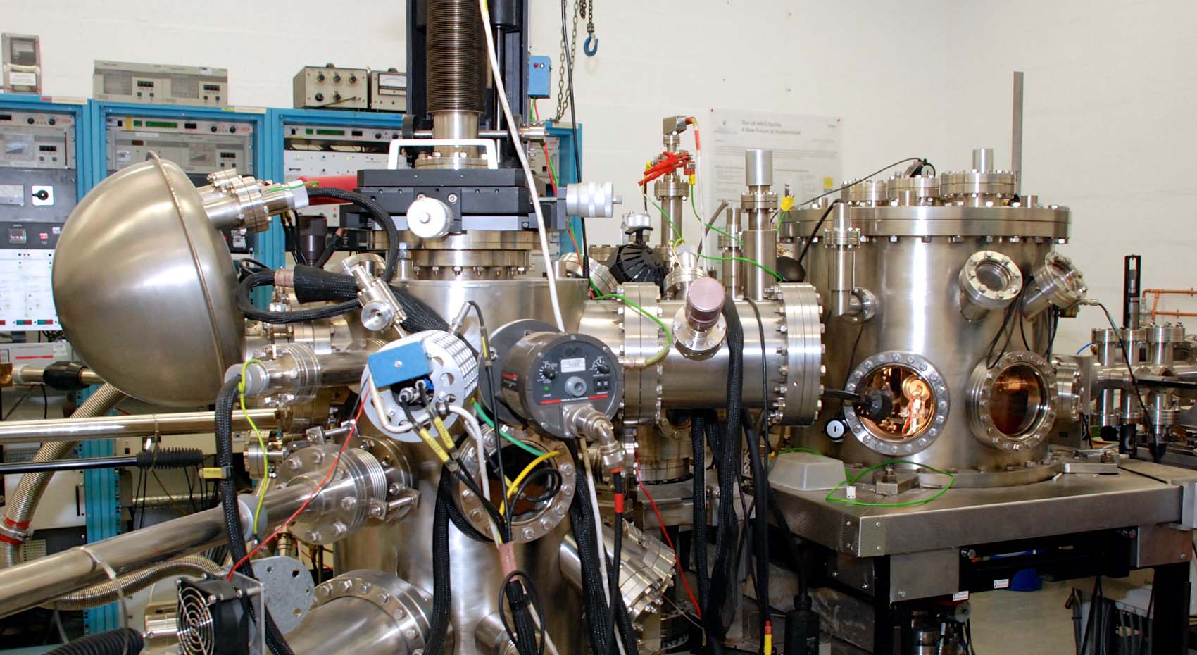
Surface Science chamber and MEIS endstation
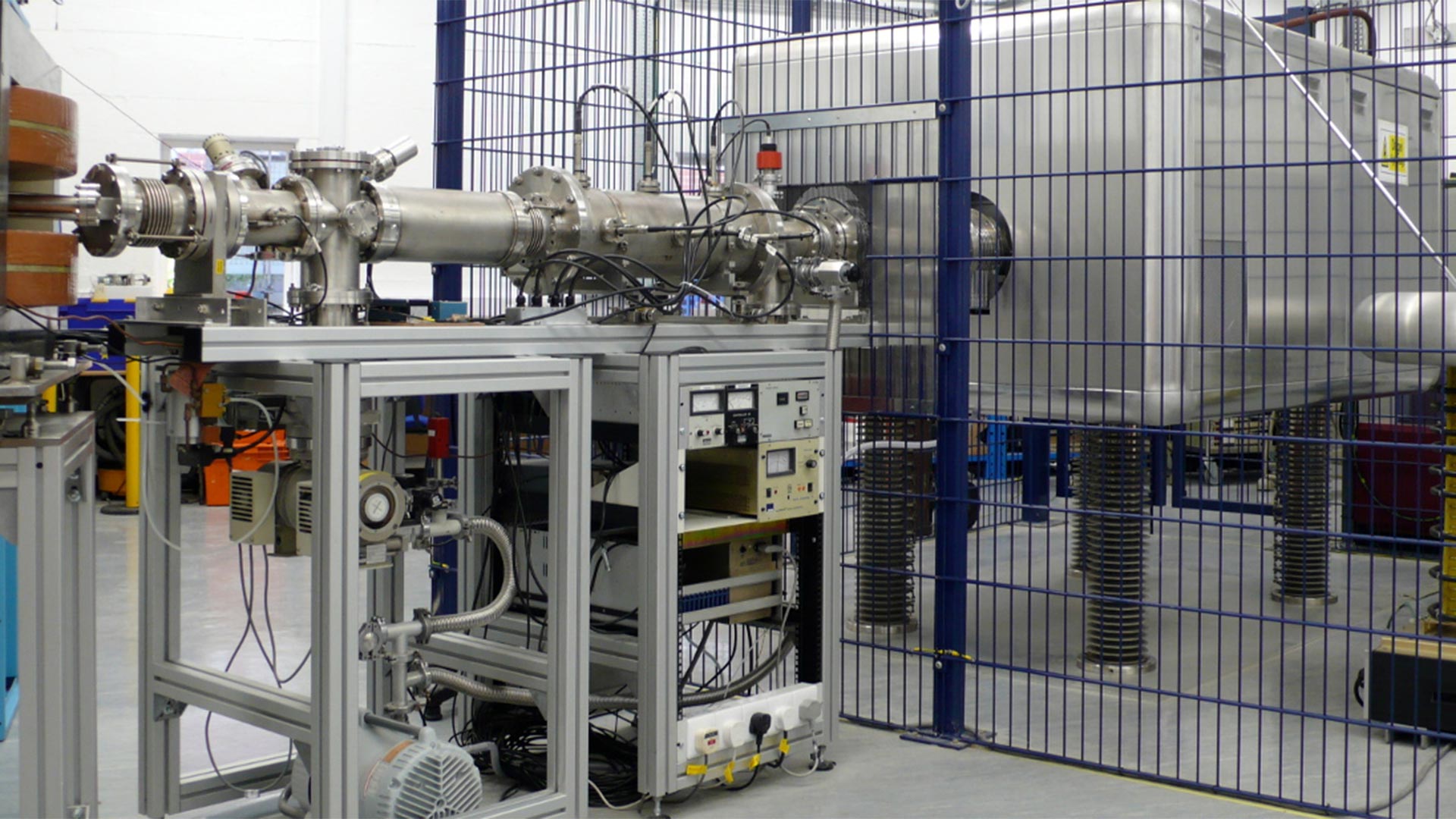
MEIS Accelerator
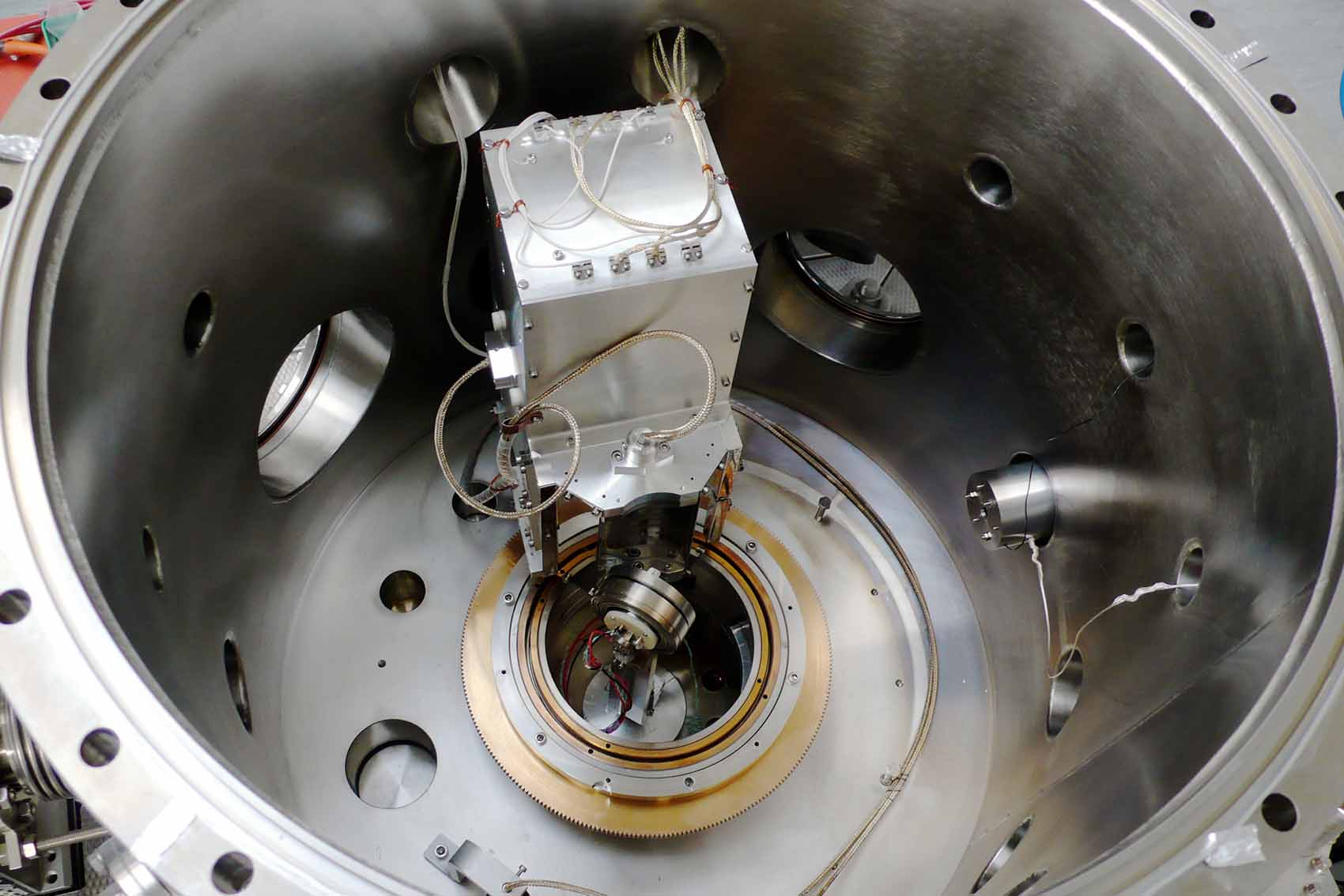
MEIS energy analyser and 3-axes goniometer
MEIS 3-axes goniometer close-up
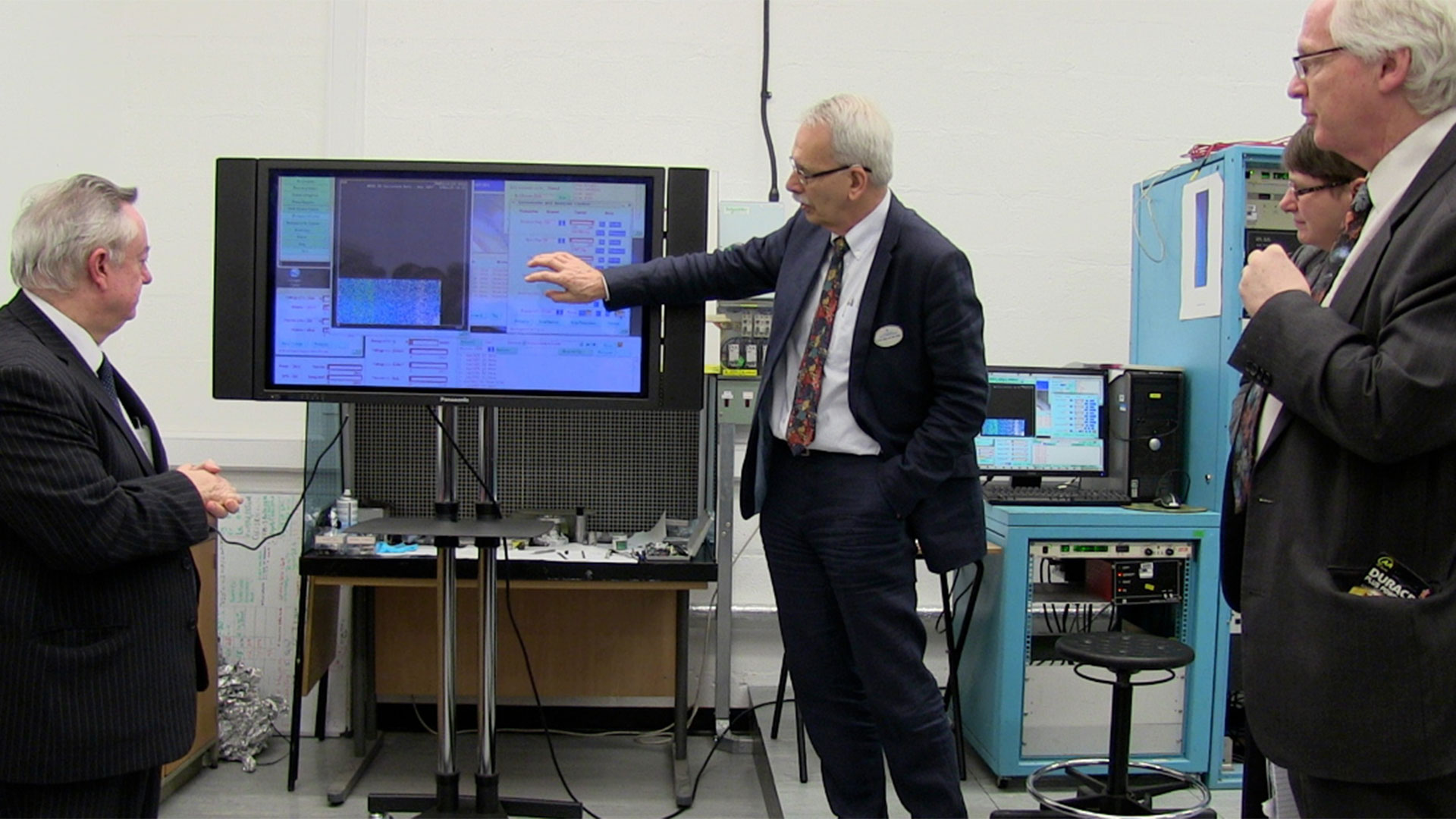
Demonstrating MEIS spectrum acquisition
Technical specification
Ion beam
- Energy range; 50-200 keV
- Species: elemental gases - typically H+ and He+
- Divergence < 0.1°
- Spot size at sample:1 mm horizontal by 0.5 mm vertical
- Current at sample: 0.1 - 1.0 μA dependent on beam energy and species
Ion Energy Analyser
- Type: toroidal electrostatic
- Analysing energy: (pass energy) 0-400 keV
- Analyser angular movement: 140° in horizontal plane
Two-dimensional (energy and angle) ion detector
- Chevron array anode with micro channel plates
- Energy window: 2% of analyser pass energy
- Resolving power: ΔE/E = 2.4x10⁻³
- Angle window: 27°
- Angular resolution: φ = 0.3°
Sample
- Size:minimum 5x5 mm, maximum 15x15 mm diameter
- Temperature range: 300-1300 K
- Environment: UHV
- Alignment: goniometer with 3 rotational and 3 translational degrees of freedom
- Loading time from air: typically 30 minutes
Preparation facilities
- Sputter cleaning
- Annealing to 1300 K
- Low Energy Electron Diffraction (LEED)
- Electron Auger
- Gas dosing
- Spare ports for evaporators and k-cells
- Sample transfer under UHV